Different Parts Of The Eye And How Does It Work
The eye is the most crucial and intricate sensory organ in the human body. The ability to visualize objects and see lights, colors and depth of what is in front of us is made possible by this vital organ.
Each eye continuously changes the amount of light it allows in (helped by the pupil), focuses on far, near and intermediate objects, and creates constant images that are immediately communicated to the brain.
Sometimes, we experience eye problems that require proper care and attention to get back to normal. So, if you currently have vision problems, we advise you to contact the Best Eye Hospital in Jakkur, Bengaluruand have your eyes examined.
How does the EYE work?
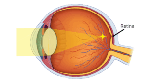
The eye is sought of a camera. When you look at something, light reflected from it enters your eyes through the pupil and is focused by the optical components within your eye.
Light enters the eye and is focused on the macula, a small area in the centre of the retina. Your ability to see fine details and colour, read, and recognize faces is all due to the macula.
Messages are transmitted from the retina to the brain via the optic nerve when light stimulates those cells. The optic nerves from the two eyes connect inside the brain. The brain uses information from each optic nerve to merge the vision from the two eyes, allowing you to see a single image.
Your vision is crucial. Often neglected, eye care should be taken into consideration or you might be at risk of permanent blindness. Consult Jakkur,Bengaluru’s Finest Eye Hospital for quality treatment right away, if you start noticing any signs or symptoms.
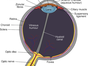
Anatomy of the Eye
This Biological Camera is made up of –
Cornea is a transparent dome-shaped surface covering the front of the eye which sends light into the eye and focuses (i.e., makes it sharp or clear).
Iris is the pigmented part of the eye that helps in adjusting the amount of light that enters it. The iris closes the pupil in order to block out strong light. In addition, the iris widens the pupil to let in more light in the night.
Pupil is the opening in the centre of the iris through which light enters the eye.
Ciliary Body is a part of the eye that produces aqueous humour.
Lens: Also known as a crystalline lens, lenses are an important part of the anatomy of eyes. The lens is positioned behind the iris. The lens directs light onto the retina by changing its shape. The lens becomes thicker to concentrate on nearby objects and thinner to focus on distant objects by the action of small muscles.
Macula is an area of the retina at the rear of the eye that is yellow and encircles the fovea that allows us to see fine details.
Fovea is the center of the macula, producing crisp and clear vision.
Retina is the layer of nerve cells inside the back of the eye that are sensitive to light. The retina detects light and generates impulses delivered to the brain via the optic nerve.
Choroid is a layer of blood vessels that lines at the back of the eye and is positioned between the retina and the sclera. In order to prevent blurry vision, it contains a pigment that absorbs extra light.
Optic nerve is a bundle of nerve fibre’s connecting the brain and retina. The visual cortex, a region of the brain, receives information from the optic nerve about light, dark, and colours and assembles it into images to produce vision.
If you’re suffering from any eye-related problem, it is crucial to get a consultation at the Best Eye Hospital in Bengaluru right away as it calls for a medical emergency.